Zydus Hospital, Vadodara, Gujarat 390020
+917015731540
drsaurabhbiswascardiologist@gmail.com
Zydus Hospital, Vadodara, Gujarat 390020
+917015731540
drsaurabhbiswascardiologist@gmail.com
Modern cardiology uses advanced tools to diagnose and treat heart conditions with high precision. IVUS (Intravascular Ultrasound), OCT (Optical Coherence Tomography), and FFR (Fractional Flow Reserve) are three powerful technologies. These help cardiologists look inside arteries, assess blockages, and decide the best treatment options.
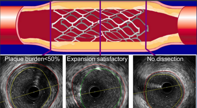
IVUS is a catheter-based imaging technique. It uses sound waves to create detailed images of the inside of blood vessels.
A tiny ultrasound probe is attached to a catheter. It’s inserted into the artery and moved to the area of interest. The device then sends out sound waves that bounce off artery walls. These echoes create real-time cross-sectional images.
IVUS is extremely helpful when angiography images are unclear.
OCT is another catheter-based imaging tool. Unlike IVUS, it uses light waves instead of sound. It offers much higher resolution and finer detail of the vessel wall.
A laser-based probe is threaded through the artery. As it moves, it captures high-definition images of the artery’s inner lining.
OCT is best used when high-resolution imaging is needed.
FFR is a technique that measures pressure differences across a coronary artery blockage. It tells doctors how much a blockage affects blood flow to the heart.
A special pressure wire is inserted into the artery. Medication is given to dilate the vessels. The wire then records pressure before and after the narrowing. The FFR value indicates if the blockage is significant.
Cardiologists often use IVUS, OCT, or FFR when:
They offer a more accurate, patient-specific approach.
| Feature | IVUS | OCT | FFR |
|---|---|---|---|
| Technology | Ultrasound (Sound Waves) | Light-based Imaging | Pressure-based Measurement |
| Resolution | Moderate | High | Not Imaging – Functional Test |
| Purpose | Artery Wall & Plaque View | High-detail Surface Imaging | Measures Blood Flow Impact |
| Best For | Stent Planning, Deep Lesions | Surface Detail, Stent Check | Assessing Functional Severity |

MBBS, MD General Medicine
DrNB Cardiology, FSCAI
Consultant Interventional Cardiologist