Contrast Echocardiography
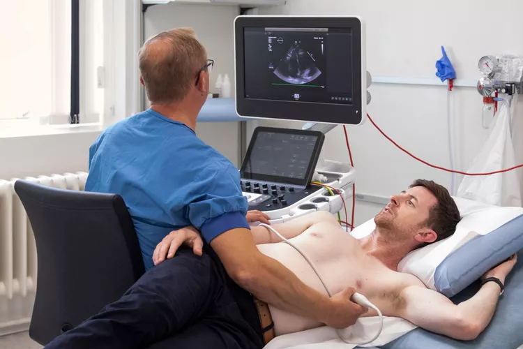
Contrast echocardiography is an advanced heart imaging technique that uses ultrasound contrast agents (microbubbles) to improve visualization of heart structures. These microbubbles are injected into a vein and enhance the echo images, especially in patients where standard echocardiography gives limited results.
It offers clearer, more detailed views of heart chambers, walls, and blood flow—crucial for accurate diagnosis.
Sometimes, standard echocardiography can’t produce good-quality images due to factors like:
- Obesity
- Lung disease
- Chest wall deformities
- Previous surgeries
In such cases, adding contrast significantly improves image clarity, making it easier for doctors to assess heart function.
How Does Contrast Work?
- A small amount of saline or contrast agent (usually perflutren lipid microspheres) is injected into a vein.
- The agent travels through the bloodstream and reaches the heart.
- Ultrasound waves bounce off the microbubbles, creating sharper and brighter images.
- The test is safe, quick, and doesn’t require anesthesia.
When Is Contrast Echocardiography Recommended?
Doctors may suggest contrast echo for:
- Poor-quality 2D images
- Evaluating left ventricular function
- Detecting clots (thrombus) in the heart
- Identifying cardiac shunts (abnormal openings)
- Assessing heart tumors or masses
- Enhancing accuracy in stress echocardiography
It's especially helpful in confirming or ruling out critical conditions like heart failure or stroke sources.
Types of Contrast Agents
- Left Heart Contrast Agents – for evaluating heart walls and function
- Right Heart Contrast (Bubble Study) – for detecting shunts like PFO (Patent Foramen Ovale)
Each has a specific use depending on the clinical need.
Benefits of Contrast Echocardiography
- Improves diagnostic accuracy
- Clarifies wall motion abnormalities
- Detects small clots missed by regular echo
- Useful in emergency and ICU settings
- Enhances confidence in cardiac reports
It allows precise interpretation even in difficult-to-image patients.
Is It Safe?
- Yes, it’s very safe for most people.
- Side effects are rare and mild
- No radiation is involved
- Allergic reactions are extremely uncommon
- It’s well-tolerated even in critically ill patients
Doctors monitor patients during and after the test to ensure safety.
Procedure Overview
- An IV line is inserted.
- The contrast agent is injected.
- Real-time imaging is captured by a trained sonographer.
- The cardiologist interprets the enhanced images.
- Results are shared soon after.