Echocardiography: 2D, 3D & Strain Imaging
Echocardiography is a non-invasive imaging test that uses ultrasound waves to create moving pictures of the heart. It helps doctors assess the heart’s structure, function, and blood flow in real time.
Echocardiography plays a crucial role in diagnosing and monitoring heart conditions, including valve disease, heart failure, congenital defects, and more.
Types of Echocardiography
1. 2D Echocardiography (Two-Dimensional Echo)
- How It Works: Produces flat, two-dimensional images of the heart, showing chambers, valves, and major vessels in real time.
- Benefits: Quick, accurate, widely available, and ideal for basic cardiac assessment. Essential for evaluating ejection fraction and valve performance.
2. 3D Echocardiography
- How It Works: Uses special probes and software to create three-dimensional views of the heart, capturing full heart volumes in one heartbeat for detailed assessment.
- Benefits: Improved visualization of valve anatomy, more accurate measurements, and critical for planning surgeries or interventions. Offers realistic views from multiple angles.
3D echo is especially useful in complex cases and pre-surgical planning.
3. Strain Imaging (Speckle Tracking Echocardiography)
- How It Works: Tracks tiny speckles on the heart muscle during each heartbeat to measure how well the heart contracts and relaxes, producing “strain” values.
- Benefits: Detects early heart dysfunction before ejection fraction drops. Useful in chemotherapy-induced cardiotoxicity, diabetic heart disease, and cardiomyopathies. Helps monitor subtle heart muscle damage in high-risk patients.
It’s a game-changer for catching heart issues before symptoms appear.
When Is Echocardiography Used?
- Chest pain or breathlessness
- High blood pressure or valve disease
- Family history of heart disease
- Undergoing chemotherapy or major surgery
- Follow-up in existing heart patients
It’s safe, painless, and doesn’t use radiation.
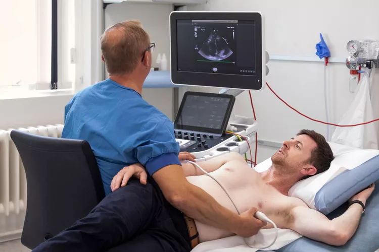
How the Test Is Performed
- You lie on a table; a gel is applied to your chest.
- A probe (transducer) sends and receives ultrasound signals.
- Images appear instantly on a monitor.
- The test lasts 20–45 minutes.
No needles, no discomfort—just real-time insights into your heart.
Advantages of Echocardiography
- Non-invasive and safe
- No radiation exposure
- Real-time imaging
- Detects both structural and functional issues
- Widely available and affordable
It’s often the first and most valuable test in cardiac evaluation.